What Is EMG? A Complete Guide to Electromyography
By ORYX Healthcare — August 2026
Every movement begins with an electrical message.
Before your hand closes, your foot takes a step, or a muscle contracts, the nervous system sends electrical signals to activate muscle fibers. Electromyography (EMG) allows clinicians to record and study this electrical activity — providing a unique window into how nerves and muscles communicate.
But what exactly is EMG? How does an EMG test work? What does the needle record, and why is electromyography so important in clinical neurophysiology?
This guide explains the fundamentals of EMG, needle electromyography, EMG signals, motor units, and nerve conduction studies in a clear and practical way.
What Is EMG?
Electromyography, commonly known as EMG, is a neurophysiological technique used to record and analyze the electrical activity of skeletal muscles.
In clinical electrodiagnostic medicine, needle EMG involves placing a small recording electrode into a selected muscle and observing its electrical activity at rest and during voluntary contraction.
The electrical signals are amplified and displayed as waveforms on an EMG system. They can also be converted into sound, allowing an experienced clinician to both see and hear characteristic patterns of muscle activity.
Although the term “EMG” is sometimes used broadly to describe an entire electrodiagnostic examination, the American Association of Neuromuscular & Electrodiagnostic Medicine (AANEM) distinguishes needle electromyography from nerve conduction studies (NCS). The two techniques provide different but complementary information.
How Do Muscles Produce Electrical Signals?
To understand EMG, it helps to understand the motor unit.
A motor unit consists of a motor neuron and the muscle fibers controlled by that neuron. When the nervous system activates a motor neuron, an electrical impulse travels along the nerve toward the muscle.
At the neuromuscular junction, this signal triggers electrical activity in the muscle fibers, followed by muscle contraction.
EMG captures these electrical events.
During voluntary contraction, multiple motor units may become active. Their electrical signals form recognizable patterns that can provide information about the functional state of muscles, peripheral nerves, nerve roots, motor neurons, and the neuromuscular system.
This is why EMG has become an important diagnostic tool in neurology and clinical neurophysiology.
How Does a Needle EMG Test Work?
During a needle EMG examination, a clinician inserts a fine needle electrode into a selected muscle.
The needle does not normally stimulate the muscle with electricity. Instead, it acts as a sensitive recording electrode that detects electrical potentials generated within the muscle.
The clinician evaluates the muscle under several conditions.
Activity During Needle Insertion
Introducing or moving the needle through muscle fibers normally produces a short burst of electrical activity known as insertional activity.
Changes in the duration or characteristics of this activity may provide useful information during the examination.
Muscle Activity at Rest
After the needle stops moving, a normal relaxed muscle is usually electrically silent, apart from activity arising from certain normal structures such as the endplate region.
Certain abnormal spontaneous potentials may appear in neuromuscular disorders.
Examples include:
- Fibrillation potentials
- Positive sharp waves
- Fasciculation potentials
- Myotonic discharges
- Complex repetitive discharges
Each pattern has specific characteristics and must be interpreted in the appropriate clinical context.
Muscle Activity During Contraction
The patient is then asked to activate the muscle gradually.
As motor units begin firing, the EMG system records motor unit action potentials (MUAPs).
Clinicians may evaluate characteristics such as MUAP duration, amplitude, configuration, firing behavior, and recruitment pattern.
As contraction becomes stronger, additional motor units are recruited and the electrical pattern becomes increasingly complex.
What Is an EMG Signal?
An EMG signal represents the electrical activity associated with muscle fiber activation.
The signal recorded by an EMG electrode depends on many factors, including the physiology of the motor unit, the location of the electrode, the distance between the recording surface and active muscle fibers, electrode geometry, and the recording system itself.
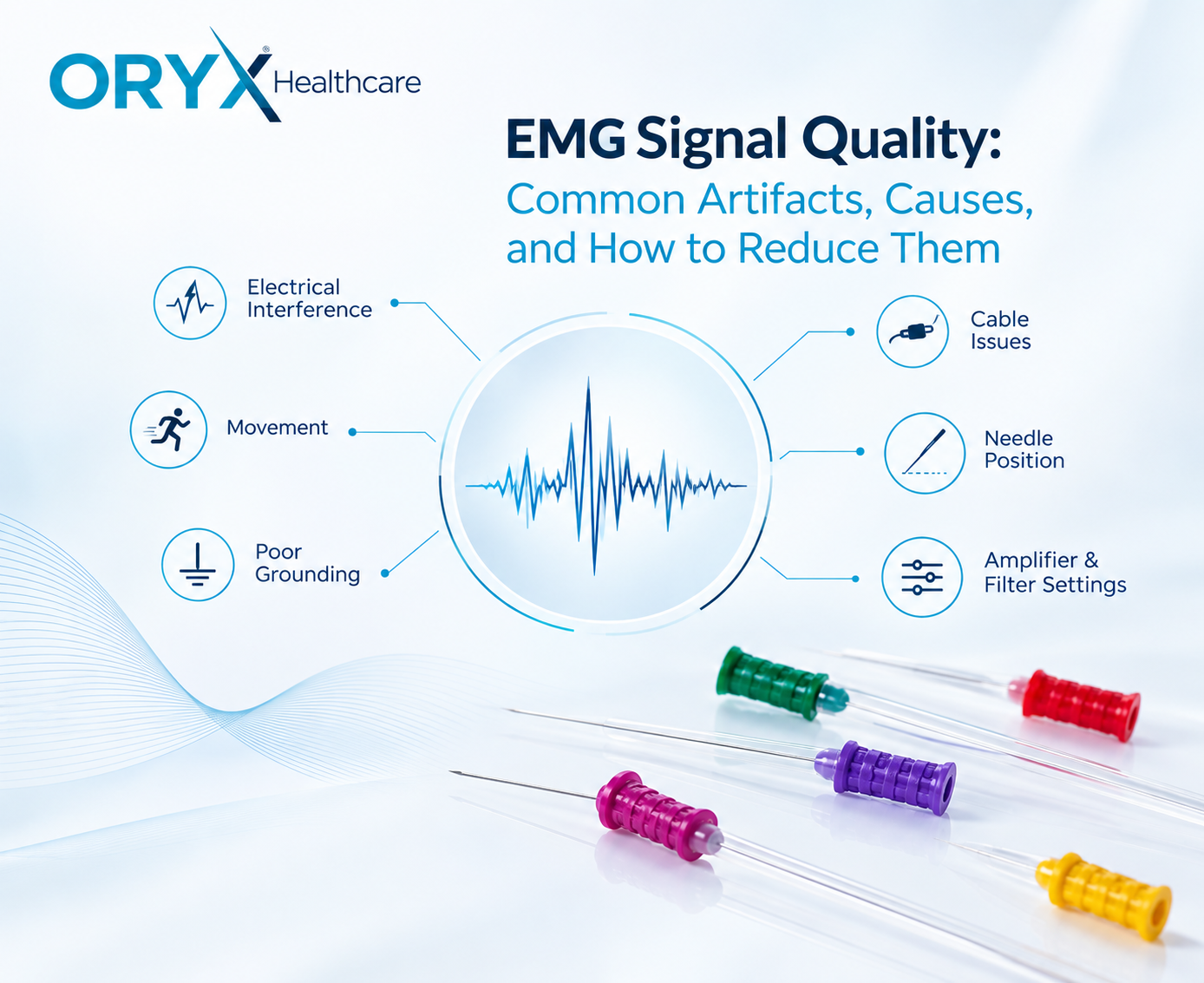
This makes electrode positioning and signal quality particularly important in needle electromyography.
The goal is not simply to detect electrical activity. The objective is to obtain a signal that can be interpreted reliably in combination with the patient’s symptoms, clinical examination, nerve conduction studies, and other diagnostic information.
What Is the Difference Between EMG and Nerve Conduction Studies?
EMG and nerve conduction studies are related, but they are not the same test.
A nerve conduction study evaluates how effectively electrical signals travel along peripheral nerves. Surface electrodes are typically used to record responses after controlled electrical stimulation of a nerve.
Needle EMG, on the other hand, records electrical activity directly from muscle.
A simple way to think about the difference is:
NCS evaluates the nerve pathway. Needle EMG evaluates electrical activity within the muscle and the motor units controlling it.
The two techniques are frequently performed together because combining their findings can help clinicians localize abnormalities within the neuromuscular system.
A detailed comparison between EMG and NCS will be explored in a separate ORYX Journal article.
What Can EMG Help Evaluate?
Electromyography can contribute to the assessment of a wide range of neuromuscular conditions.
Depending on the clinical presentation and the complete electrodiagnostic examination, EMG may provide useful information when investigating conditions involving:
Peripheral nerves
such as peripheral neuropathies and focal nerve injuries.
Nerve roots
including suspected cervical or lumbosacral radiculopathy.
Muscle
including different types of myopathy.
Motor neurons
in disorders affecting the motor neuron system.
Neuromuscular transmission
where specialized electrophysiological techniques may be used to assess communication between nerves and muscles.
EMG findings are not interpreted in isolation. A complete electrodiagnostic evaluation is guided by the patient’s history, physical examination, symptoms, and other available investigations.
Why Is the Needle Electrode Important?
An EMG system may contain sophisticated amplifiers, filters, software, and signal-analysis tools, but the recording process begins at one very small interface:
the electrode inside the muscle.
The design and electrical characteristics of an EMG needle can influence the recording environment.
Important factors include:
Recording surface area — influences the region from which muscle electrical activity is detected.
Electrode geometry — affects the spatial relationship between the recording surface and surrounding muscle fibers.
Needle positioning — even small changes in position can alter the recorded motor unit waveform.
Electrical characteristics — electrode impedance and connection quality can influence recording conditions.
Mechanical characteristics — needle diameter, length, tip geometry, and handling characteristics are relevant to clinical use.
For this reason, consistent electrode design and manufacturing are important considerations in diagnostic needle electromyography.
What Is a Concentric EMG Needle?
One commonly used electrode design in clinical needle EMG is the concentric needle electrode.
A concentric electrode contains a recording element positioned within a metal cannula. The central recording surface and surrounding reference are located close to one another.
This configuration provides a localized recording arrangement suitable for examining motor unit activity.
Concentric needle electrodes have played an important role in electromyography for almost a century. In fact, the concentric needle concept was described in early human motor-unit recordings by Edgar Adrian and Detlev Bronk in 1929.
Modern concentric EMG needles continue to be widely used in clinical neurophysiology.
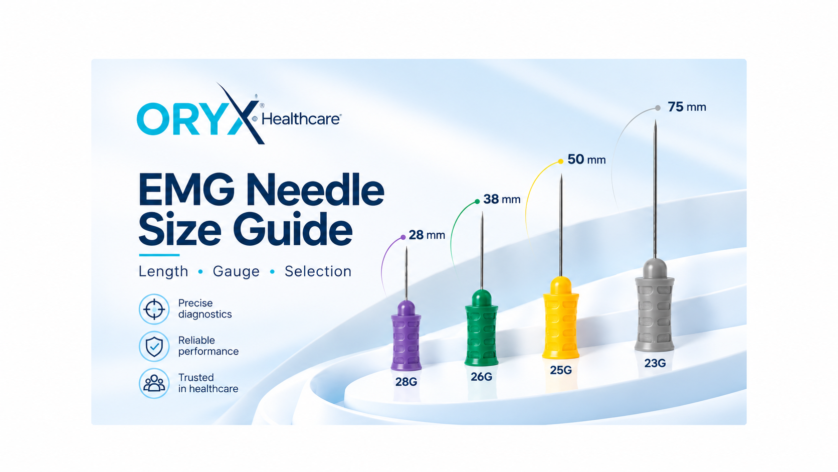
ORYX Healthcare develops disposable concentric EMG needle electrodes in multiple lengths and gauges for different clinical requirements.
Needle EMG vs Surface EMG
Not all electromyography uses an inserted needle.
Surface EMG (sEMG) records electrical activity through electrodes placed on the skin. It is widely used in areas such as biomechanics, rehabilitation, movement analysis, ergonomics, sports science, and research.
Needle EMG records activity from within the muscle and provides greater spatial selectivity for clinical assessment of individual motor unit activity.
The appropriate method depends on the purpose of the examination.
Recent developments in high-density surface EMG (HD-sEMG) have expanded the possibilities of non-invasive motor-unit analysis and neuromuscular research. However, needle EMG remains an established component of clinical electrodiagnostic medicine.
Does an EMG Test Hurt?
Because needle EMG requires inserting a small electrode into muscle tissue, patients may experience temporary discomfort during insertion or repositioning.
The sensation can vary according to the muscle examined, needle dimensions, insertion technique, individual sensitivity, and other factors.
After an examination, temporary tenderness or mild bruising may occasionally occur.
The examination should always be performed by appropriately trained healthcare professionals following relevant clinical procedures and precautions.
What Does an Abnormal EMG Mean?
There is no single waveform that simply means “abnormal EMG.”
The interpretation depends on the complete pattern observed during the examination.
Clinicians consider multiple findings, including spontaneous activity at rest, motor unit action potential characteristics, recruitment, distribution of abnormalities across muscles, and nerve conduction results.
The same individual waveform may have different significance depending on the clinical situation.
For this reason, EMG should be viewed as an extension of the neurological and neuromuscular examination rather than as an isolated laboratory result.
EMG Is Entering a New Technological Era
Electromyography continues to evolve.
Modern research is exploring advanced signal processing, high-density electrode systems, automated motor-unit decomposition, quantitative EMG analysis, and artificial intelligence.
A 2024 review published in Clinical Neurophysiology examined the use of artificial intelligence for automatic classification of needle EMG signals. Research has also investigated AI-based classification of motor unit action potentials in real-world needle EMG recordings.
At the same time, high-density surface EMG is creating new possibilities for non-invasive analysis of motor-unit behavior.
These technologies may eventually provide clinicians with additional quantitative tools, but interpretation of electrophysiological findings remains closely connected to clinical knowledge and neurophysiology expertise.
Why EMG Still Matters
Almost a century after the first human recordings with concentric needle electrodes, EMG remains one of the most direct ways to observe the electrical behavior of the neuromuscular system.
It transforms tiny electrical signals inside muscle into information that can help clinicians understand how nerves, motor units, and muscles are functioning.
And despite increasingly sophisticated technology, the principle remains remarkably elegant:
listen to the electrical language of muscle.
Frequently Asked Questions About EMG
What does EMG stand for?
EMG stands for electromyography, a technique used to record and analyze the electrical activity of skeletal muscles.
What does an EMG test measure?
Needle EMG records electrical activity generated by muscle fibers and motor units when the muscle is at rest and during voluntary contraction.
Is EMG the same as a nerve conduction study?
No. Needle EMG records electrical activity within muscles, while a nerve conduction study evaluates electrical signal transmission along peripheral nerves. They are frequently performed together.
What type of needle is used for EMG?
Several needle electrode designs exist. Concentric needle electrodes are commonly used in clinical electromyography.
What is a motor unit action potential?
A motor unit action potential, or MUAP, is the electrical potential recorded from muscle fibers belonging to a motor unit. Its characteristics are evaluated during needle EMG.
Can EMG diagnose neurological disorders?
EMG can provide important information in the assessment of neuromuscular and neurological conditions, but results must be interpreted together with the patient’s clinical history, physical examination, nerve conduction studies, and other diagnostic findings.
References
- American Association of Neuromuscular & Electrodiagnostic Medicine (AANEM). Overview of Electrodiagnostic Medicine. Position Statement.
- MedlinePlus, U.S. National Library of Medicine. Electromyography (EMG) and Nerve Conduction Studies.
- Farina D, Gandevia S. The neural control of movement: a century of in vivo motor unit recordings is the legacy of Adrian and Bronk. The Journal of Physiology. 2024;602(2):281–295. DOI: 10.1113/JP285319.
- de Jonge S, Potters WV, Verhamme C. Artificial intelligence for automatic classification of needle EMG signals: A scoping review. Clinical Neurophysiology. 2024;159:41–55. DOI: 10.1016/j.clinph.2023.12.134.
- Shandiz E, et al. Applying high-density surface EMG to the study of neuromuscular disorders: a systematic review. Clinical Neurophysiology. 2025.
Medical information disclaimer: This article is intended for professional and educational information and does not replace medical diagnosis, clinical judgment, or individual medical advice.