EMG vs. NCS: What’s the difference, and why are these two tests so often performed together?
Electromyography (EMG) and nerve conduction studies (NCS) are two fundamental tools in electrodiagnostic medicine. Although both tests help clinicians evaluate the neuromuscular system, they examine different parts of the nerve–muscle pathway. While NCS primarily assesses how effectively electrical signals travel through peripheral nerves, EMG evaluates the electrical activity within muscles. Therefore, using these two tests together can provide a more complete picture of neuromuscular function and help clinicians identify the source of a patient’s symptoms more accurately.
In simple terms:
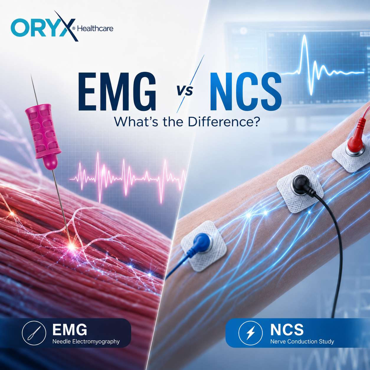
NCS evaluates how electrical signals travel along peripheral nerves.
Needle EMG records and evaluates electrical activity within skeletal muscle.
Because nerve and muscle function are closely connected, the two tests often provide complementary information.
What Is an EMG Test?
Electromyography, or EMG, is used to record and analyze electrical activity generated by skeletal muscles.
During needle EMG, a fine recording needle electrode is inserted into selected muscles.
The examiner evaluates electrical activity during different stages of the examination, including:
- Needle insertion
- Muscle relaxation
- Minimal voluntary contraction
- Increasing muscle contraction
The recorded activity can provide information about motor units and the nerves controlling the examined muscle.
Important findings may include:
- Insertional activity
- Spontaneous activity
- Motor unit action potentials
- Motor unit firing behavior
- Recruitment patterns
What Is a Nerve Conduction Study?
A nerve conduction study, commonly abbreviated as NCS, evaluates how electrical signals travel through peripheral nerves.
During the examination, a controlled electrical stimulus is applied to a nerve while recording electrodes measure the resulting response.
Depending on the nerve being studied, clinicians may evaluate parameters including:
- Conduction velocity
- Response amplitude
- Distal latency
- Waveform morphology
- Temporal dispersion
- Conduction block
These measurements provide information about sensory and motor nerve function.
EMG vs NCS: What Is the Main Difference?
The key difference is where the electrical activity is being evaluated.
NCS Examines the Nerve
Nerve conduction studies evaluate the transmission of an electrical signal through a peripheral nerve.
Changes in conduction speed, response amplitude, or waveform characteristics may provide information about nerve dysfunction.
Needle EMG Examines the Muscle
Needle EMG records electrical activity directly from skeletal muscle.
Because muscles are controlled by motor neurons, changes in muscle electrical activity can provide information about the motor unit and the nerve supplying the muscle.
EMG vs NCS at a Glance
| Feature | Needle EMG | Nerve Conduction Study |
|---|---|---|
| Main area evaluated | Muscle and motor units | Peripheral nerves |
| Recording method | Needle electrode | Usually surface electrodes |
| Electrical stimulation | Not normally delivered through the recording needle | Nerve is electrically stimulated |
| Motor unit analysis | Yes | No |
| Recruitment analysis | Yes | No |
| Conduction velocity | No | Yes |
| Often part of the same EDX examination | Yes | Yes |
Why Are EMG and NCS Often Performed Together?
The neuromuscular system is a connected pathway.
Symptoms such as:
- Weakness
- Numbness
- Tingling
- Muscle wasting
- Cramping
- Pain
may result from abnormalities at different locations.
The problem could involve:
- A peripheral nerve
- A nerve root
- A plexus
- A motor neuron
- The neuromuscular junction
- Skeletal muscle
One technique may not provide all the information required to localize the problem.
NCS provides information about peripheral nerve conduction, while needle EMG provides information about muscles and motor units.
Together, they can help clinicians determine where along the nerve–muscle pathway an abnormality may be located.
What Can a Nerve Conduction Study Reveal?
Nerve conduction studies are especially useful in the evaluation of peripheral nerve disorders.
For example, NCS can contribute to the assessment of:
- Focal mononeuropathies
- Polyneuropathies
- Entrapment neuropathies
- Demyelinating processes
- Axonal nerve injury
Patterns involving amplitude, latency, conduction velocity, and waveform characteristics may help characterize the nature of peripheral nerve dysfunction.
However, results must always be interpreted in the context of the patient’s clinical presentation.
What Can Needle EMG Reveal?
Needle EMG provides a different type of information.
When a muscle is at rest, the examiner evaluates whether abnormal spontaneous electrical activity is present.
During voluntary contraction, the examiner evaluates motor unit action potentials (MUAPs) and recruitment.
These findings may contribute to distinguishing patterns associated with:
- Neurogenic disorders
- Myopathic disorders
- Denervation
- Reinnervation
- Chronic motor unit remodeling
Research continues to improve quantitative interpretation of needle EMG. Recent work has established reference values for motor unit recruitment in commonly examined muscles, demonstrating that even established EMG methods continue to evolve.
EMG vs NCS in Radiculopathy
Radiculopathy is a useful example of why EMG and NCS provide complementary information.
A nerve-root lesion is located proximal to the dorsal root ganglion. As a result, sensory nerve conduction studies may sometimes remain normal despite clinically significant radiculopathy.
Needle EMG can provide additional information by examining muscles supplied by the suspected nerve root.
The combination of clinical examination, NCS, and needle EMG can therefore be useful in localizing the lesion.
EMG vs NCS in Carpal Tunnel Syndrome
Nerve conduction studies play an important role in evaluating suspected carpal tunnel syndrome, where the median nerve is compressed at the wrist.
NCS can examine conduction across the affected nerve segment.
Needle EMG may also be included depending on the clinical question and findings.
The appropriate combination of tests is determined by the clinician performing the electrodiagnostic evaluation.
Does NCS Use a Needle?
Standard nerve conduction studies generally use electrodes placed on the skin.
The electrical stimulation patients feel during the examination is normally part of the nerve conduction study.
Needle EMG is different: a small needle electrode is inserted into selected muscles to record their electrical activity.
Does the EMG Needle Electrically Stimulate the Muscle?
In conventional needle electromyography, the needle primarily functions as a recording electrode.
It detects electrical potentials produced by muscle fibers.
This is different from nerve conduction studies, in which electrical stimulation is intentionally applied to a peripheral nerve.
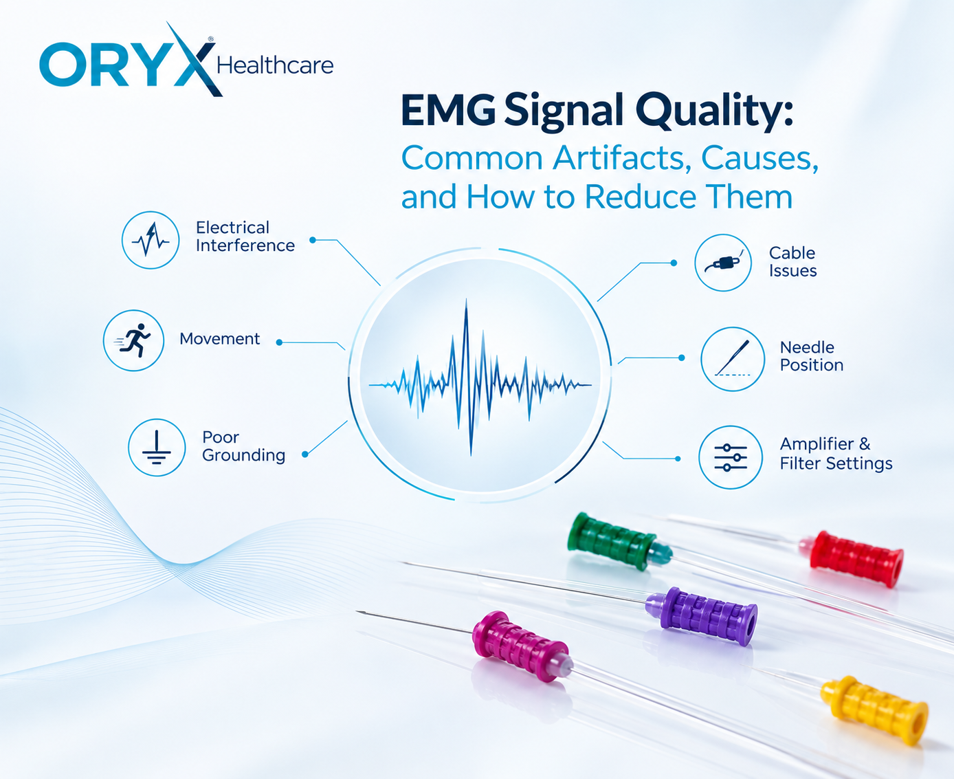
Why Electrode Quality Matters
Electrodiagnostic signals can be small and sensitive to technical factors.
Reliable recordings depend on factors including:
- Electrode positioning
- Stable electrical connections
- Recording environment
- Skin preparation for surface electrodes
- Cable integrity
- Grounding
- Electrode characteristics
In needle EMG, the recording electrode forms the direct electrical interface with the muscle.
Consistent electrode geometry and electrical characteristics are therefore important parts of the recording chain.
ORYX Healthcare develops disposable concentric EMG needle electrodes for clinical needle electromyography.
Two Tests, One Neuromuscular System
A nerve carries the command.
A muscle produces the response.
NCS examines electrical conduction along the nerve.
Needle EMG examines electrical behavior within the muscle.
Rather than asking which test is “better,” it is more useful to understand the different information each provides.
EMG and NCS are two complementary views of the same neuromuscular system.
Frequently Asked Questions
Is EMG the same as NCS?
No. Needle EMG records electrical activity inside skeletal muscle, while NCS evaluates electrical signal conduction along peripheral nerves.
Why are EMG and NCS usually performed together?
They provide different but complementary information that can help localize and characterize abnormalities in the neuromuscular system.
Does NCS use needles?
Standard nerve conduction studies generally use surface recording electrodes. Needle electrodes are used during needle electromyography.
Which test measures nerve conduction velocity?
Nerve conduction studies measure conduction velocity. Conventional needle EMG does not.
Can EMG and NCS diagnose every nerve or muscle disorder?
No single electrodiagnostic test can diagnose every condition. Results are interpreted together with the patient’s history, clinical examination, and other diagnostic information.
References
- American Association of Neuromuscular & Electrodiagnostic Medicine (AANEM). Types of Tests: Needle EMG and Nerve Conduction Studies.
- American Association of Neuromuscular & Electrodiagnostic Medicine (AANEM). Reporting the Results of Nerve Conduction Studies and Needle EMG.
- Hannaford A, Paling E, Silsby M, et al. Electrodiagnostic studies and new diagnostic modalities for evaluation of peripheral nerve disorders. Muscle & Nerve. 2024;69(6):653–669. DOI: 10.1002/mus.28068.
- Rubin DI, Lamb CJ. Motor unit potential recruitment reference values in common upper and lower extremity muscles. Clinical Neurophysiology. DOI: 10.1016/j.clinph.2024.05.001.
Medical information disclaimer: This article is intended for professional and educational purposes and does not replace medical diagnosis, clinical judgment, or individual medical advice.